Robotic Knee & Hip Replacement, Shoulder & Sports Surgery
Robotic precision for hip & knee replacement. Computer-navigated, customised shoulder replacement. Arthroscopic (keyhole) surgery for ligament, cartilage, meniscal and shoulder problems — by one of Delhi NCR's most trusted orthopaedic surgeons.
Book Your Appointment
Quick callback — share your details and our team will reach out shortly.
Trusted hands. Robotic precision.
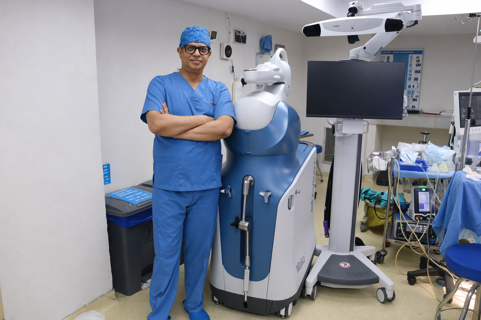
Dr. Dhananjay Gupta
Senior Consultant — Robotic Joint Replacement & Orthopaedic Surgery
Dr. Dhananjay Gupta is among Delhi NCR's most experienced robotic joint replacement surgeons, specialising in robotic-assisted knee, hip and shoulder surgeries. He has performed over 15,000 successful joint surgeries with a strong focus on minimally invasive techniques, faster recovery and long-lasting outcomes.
- MBBS, MS (Orthopaedics), Fellowship in Joint Replacement
- 30+ years of orthopaedic experience
- 15,000+ successful joint surgeries performed
- Specialist in robotic knee & hip replacement, navigated shoulder replacement, and arthroscopic sports surgery
- Featured at leading hospitals across Delhi NCR
Robotic, Navigated & Arthroscopic Care
Robotic precision for hip & knee replacement; navigated, customised replacement for the shoulder; arthroscopic surgery for ligaments, cartilage, meniscus and shoulder problems — comprehensive joint care under one expert.
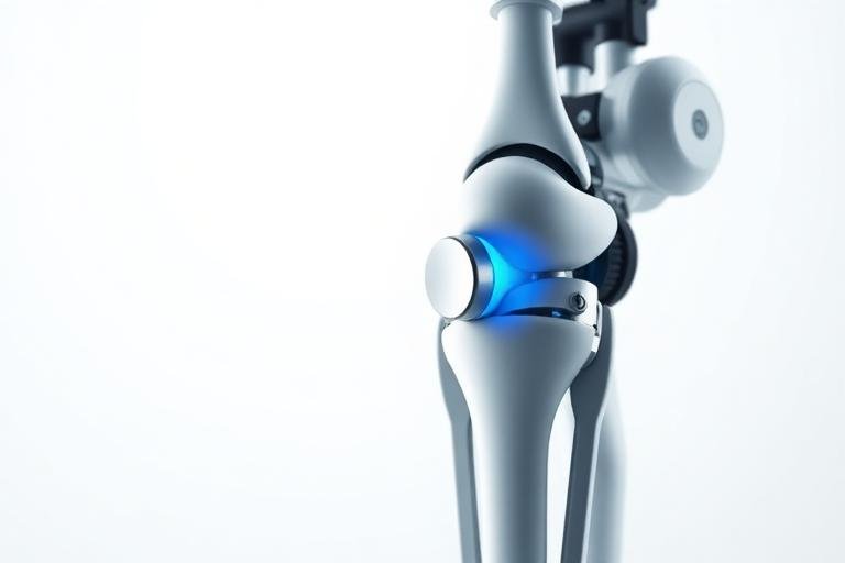
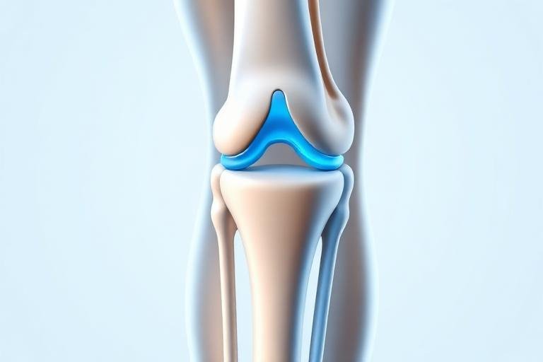
Robotic Knee Replacement
AI-guided, sub-millimetre precision knee replacement with faster recovery and natural movement.
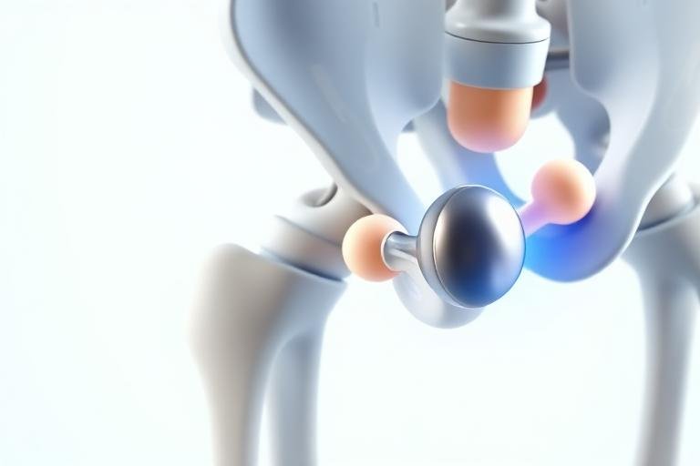
Robotic Hip Replacement
Minimally invasive robotic hip replacement that restores mobility with smaller incisions.
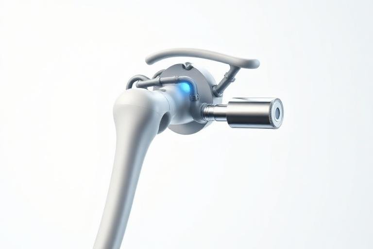
Shoulder Surgery
Navigated & customised shoulder replacement plus arthroscopy for frozen shoulder, dislocation, rotator cuff, impingement and SLAP/PASTA lesions.
Ligament, Cartilage & Meniscus Repair
Arthroscopic ACL/PCL and multi-ligament reconstruction, cartilage repair and meniscal repair to get athletes and active patients moving again.
Arthroscopic Surgery
Day-care keyhole surgery for the knee, shoulder and elbow — minimal scarring, fast recovery, no robot.
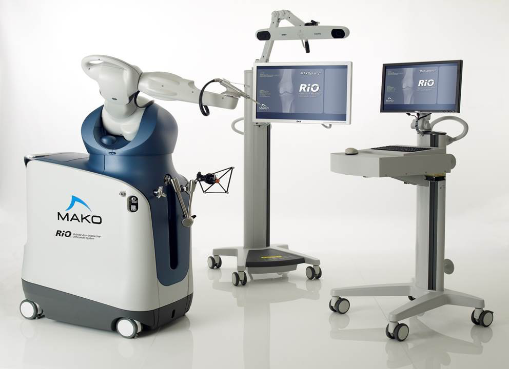
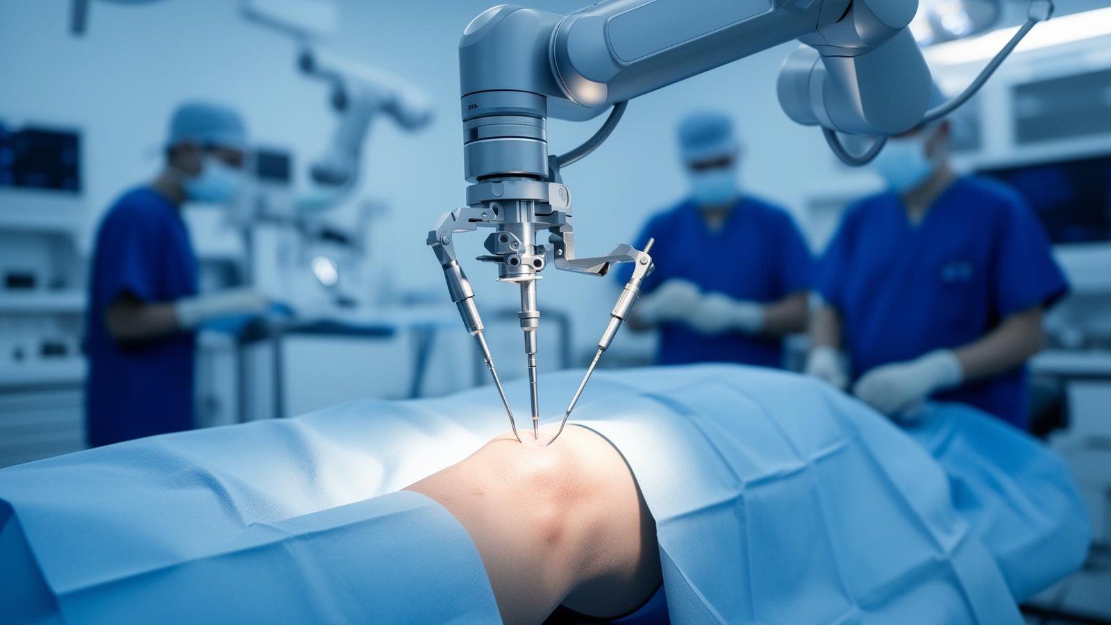
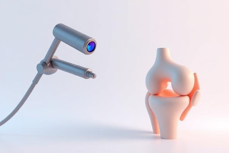
Powered by next-generation surgical robotics
Every implant is positioned with sub-millimetre accuracy using real-time 3D imaging — for joints that feel and move naturally.
Real recoveries. Real results.
Hear from patients who have returned to a pain-free, active life after surgery.
"I was bedridden with knee pain for two years. Three weeks after my robotic surgery, I was walking 5 km a day. Best decision of my life."
"The doctor and his team explained everything clearly. Surgery was painless and recovery was much faster than I expected."
"I tore my ACL playing cricket. Today, six months after arthroscopic surgery, I'm back on the field stronger than before."
Frequently Asked Questions
Clear, doctor-approved answers to the questions patients ask us most.
Start Walking Pain-Free Again
Book a consultation with Delhi NCR's leading robotic orthopaedic surgeon. Same-week appointments available.